Weight Lifting Injuries

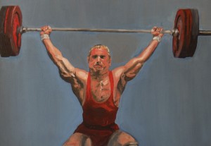
Statistically, more and more people are working out using weights and weight bearing exercise than ever before.  Whether it’s to achieve those major biceps for men or to tone and stave off the ravages of osteoporosis for women,  fitness with weights is at all an all time high.  So, unfortunately, are serious shoulder injuries, most of which occur with upper arm strengthening.

One common problem  is an injury to the A-C joint  The A-C Joint is the small accessory joint at the top of the shoulder. You can feel it — it is the small bump at the end of your collarbone. A-C stands for Acromio-Clavicular. The acromion is a flat bone at the top end of the shoulder blade. It is nature’s shoulder-pad — a square shaped bone that you can feel at the top of your shoulder.  The clavicle is the collarbone. Between the end of the clavicle and the acromion is the A-C joint which plays a major supporting role in the function of the shoulder.”

Both those who work out and their fitness trainers can stress the A-C joint when they do bench-presses and overheads because any exercise that uses a substantial amount weight to move the arm also involves stress to the shoulder.  Both of these lifts strengthen the powerful deltoid muscle which lies along the acromion and the clavicle and thus, the stress of heavy lifting can be transmitted to the small A-C joint. People who lift weights for years may injure the A-C joint and develop arthritic degeneration in the area. My patients frequently complain of pain and popping  at this joint.   I can even hear the pop on examination before x-ray or MRI confirms the injury or arthritic condition.”

The bad news on A-C  joint degeneration is that  once the damage occurs, it cannot be reversed. There is no cure for arthritis. One solution might be to curtail or give up overhead lifting, but this is usually not an option for dedicated weight lifters.  They may abstain from or limit weight lifting, but usually only for short periods of time and rarely long enough to stabilize a serious joint condition. Anti-inflammatory medicines like Aleve, Celebrex and Advil  may relieve pain but their effects may only be temporary.  A cortisone injection into the A-C  joint may also help, albeit temporarily. And neither will relieve the ‘popping’ of the joint.  That said, both therapies  should be tried prior to surgical intervention.

When A-C joint surgery is indicated, both the pain of the injury and the ‘popping’ sound can be cured with comparatively minor surgery. Quite simply, the end of the collarbone is removed. The painful arthritis and the pressure at the joint are relieved. This often ends both the pain and the popping.  The surgery can be done either arthroscopically or through a very small incision at the top of the shoulder. Recuperation is relatively quick  usually two to three weeksto return to normal activity .  The good news for the gym set is that there is usually little or no postoperative impairment to the shoulder and patients can happily resume weight lifting and other sports. Some lifters may note slight loss of maximum bench press strength, but this will be offset by relief of painful popping.

Weight lifters can also develop muscular injuries from overuse or overdoing it! One common condition is bicepital tendinitis.  “The biceps muscle bends the elbow and it’s long tendon lies in a groove in the front of the shoulder. Tendinitis or inflammation of the tendon can occur with bench presses and ‘dips’.  Treatment consists of pain and/or anti-inflammatory medication ; Physical therapy can be useful.  Sometimes, a cortisone injection. is necessary

Older lifters may even rupture the tendon. Sharp pain and a popping sensation may be felt in the front of the shoulder. The pain will eventually subside and usually there will be little or no lasting disability so surgery is rarely necessary.

Another muscle that can tear with excessive stress during bench presses is a tear to the so-called “pecs” – the pectoralis major muscles. A partial tear can be treated with rest and therapy and should heal on it’s own. A complete tear may require surgical repair.

Working out with weights is an important part of many people’s lifestyles. It is, for the most part, an excellent way to help stay fit and an anti-aging boost.  That  said, it must be approached with care in order for people to gain the maximum benefit from work-outs.”  In short, take care of your arms and shoulders and they will help take care of you.  If not, your upper body fitness is at serious and potentially permanent risk due to injury.

 Please note: these articles are for general information. They are not intended to serve as medical advice or treatment for a specific problem. Diagnosis and treatment of a problem can only be accomplished in person by a qualified physician.
Runner’s Knee

Chondromalacia, refers to a condition in which there is damage to the joint surface of the patella (kneecap).  It is common in runners, cyclists, catchers –any athlete that is constantly bending their knees.  At a large runner’s clinic several years ago, knee problems predominated, and the most common cause of the pain was patella disease.

Runners with chondromalacia complain of a deep ache under the patella.  It is present during running and is aggravated by jogging up and down hills.  Frequently, the runner will complain of a noisy knee—a crackling sound is heard and felt.  Running through this injury only produces more pain and swelling.

CAUSE

The cause of the pain and the noise is a roughness on the underside of the patella.  Normally, the joint surface of the patella is very smooth—like a slick piece of ice.  Damage to the patella produces scratches and nicks so that eventually the surface becomes rough and irregular. Think of an ice skating rink after a hockey game.  As the knee bends, the patella is pressed tighter and deeper into the knee, and this causes pain, just as rubbing a skin wound would.  The knee becomes inflamed, and swelling can result.

Why does the damage occur?  Normally, the patella moves in a straight line as the knee bends and straightens. The patella is wedge-shaped and lies in a bony groove on the top of the femur (thigh bone).  This groove holds it in proper alignment.  If an abnormality is present to disrupt this alignment, the patella can slip out of the groove as it moves, and a shearing damage to its surface can occur.  This injury may be minor each time, but over the years the damage becomes extensive.  The most common abnormality is a combination of a “knocked knee” posture of the leg and tightness of the ligaments on the outer side of the knee.  A bowstring effect occurs and pulls the patella out of line.  Other causes include deformities of the patella or shallowness of the groove. Abnormalities of the foot, and even the hip can contribute. For example ,intoeing from the hip joint or pronation of the feet can lead to patella abnormalities and can influence proper patella alignment and tracking.

Any direct injury to the patella, such as a fall or fracture, can also produce this problem.  Athletes, such as baseball catchers or football linemen, who must constantly squat or kneel can also be susceptible.

TREATMENT

As one would imagine, treatment consists of rest from running and avoidance of excessive bending.  Since hill running produces constant knee flexion, changing to a flat surface, such as a track, is advisable.  Anti-inflammatory medication is useful for this condition and can be taken for the pain.  (It is best to consult your doctor before taking frequent doses.)

The most important factor in treatment, however, is a good exercise program.  To counter the tight bowstringing ligaments which pull on the outer side, the muscles on the inside of the knee must be developed and strengthened.  The best exercise is performed while lying flat with the knee completely straight.  The leg is lifted up and held to the count of ten.  During this time, an isomeric tightening of the knee muscles can also be done.  Lower the leg, rest for a few seconds, and raise it again.  One should gradually work up to 100-150 lifts per day (in intervals).  Ankle weights can also be used starting with two pounds and increasing to ten pounds over several weeks.  Bending exercises should generally be avoided because they increase the pressure on the patella and thereby aggravate the condition.

Later, when the pain subsides, running can be resumed with a graduated program.  Ice should be applied after the run.  Knee exercises should continue and hills avoided.

If the problem persists, various knee bands or braces can be prescribed.  The most common type of brace for chondromalacia has a hole over the kneecap to hold it in place and to prevent abnormal side displacement.  If the runner has pronated feet, a shoe orthotic may help to better position the heel and take pressure off of the knee.

In severe cases of chondromalacia, the patella may partially dislocate (sublux), producing further damage and disability.  Conservative medical measures already discussed should first be initiated; but if they fail, surgery is warranted.  Operative procedures can be performed with the arthroscope through small incisions.  The tight lateral ligaments can be cut ( lateral release), and the rough surfaces of the patella can be shaved and smoothed to some extent.  This operation is frequently successful.

I remember back to my training just prior to the advent of arthroscopic surgery. Operations on the patella for this conditions consisted of an open incision to re-align the patella. Tissues were cut on one side of the knee and others, on the other side were tightened. The results were frequently dismal. Now with arthroscopic surgery, the results are much better- achieved with minor surgery. This has been a great advance in medicine.

True dislocations of the patella can occasionally occur—usually as the result of trauma. They are usually treated with a brace to allow the torn ligaments to heal. Recurrent dislocations may require surgery. Sometimes, an open reconstruction: the results are not always uniform. Therefore conservative measures or even arthroscopic surgery should first be tried.

To conclude, in all of these injuries, prompt recognition and treatment, as well as thoughtful medical and technical measures, is important to prevent permanent impairment.

Elbow Pain

In this article I discuss elbow pain relating to both baseball and tennis. If you play other sports that involve throwing and are experiencing elbow pain, refer to the baseball section of this article. If you are playing other racket sports, refer to the tennis section.

Throwing Related Injuries

Major league and Little League pitchers have one thing in common:  they frequently develop pain along the inner side of the elbow.

The pain is caused by throwing, and therefore it is important to understand the mechanism and to be able to recognize and manage various injuries.    As the arm accelerates forward, the muscles along the inner side of the elbow become very active and stretch.

Four Phases of Throwing:

  1. Stance Phase.  The body is relaxed.
  2. Windup.  The shoulder lifts and rotates outward as the arm is cocked.  The muscles in front of the shoulders and around the elbow tighten.  Because of increased tension, muscle injuries can occur.  The muscles in front of the shoulder can be sprained, and the biceps tendon in the shoulder can become inflamed.
  3. Acceleration or forward motion.  Considerable force is generated across the shoulder and elbow during this short phase.  The shoulder moves forward and swings from outward to internal rotation producing a rather violent torque at the ball of the shoulder.  There is little active shoulder muscle contraction:  the arm is hurled forward by trunk rotation and trunk muscles.  Still, rotatory force at the shoulder joint may produce muscular injuries in adults and fractures in children.

The muscles along the inside of the elbow are very active during this phase and are tensed and stretched.  If the elbow is overused and abused, inflammation, pain, and tenderness result.

  1. Follow-through.  As the ball is released, there is an increase of shoulder activity.  Also, the powerful triceps muscle behind the arm contracts to straighten the elbow.  The forearm and wrist rotate if a curve of slider is to be thrown.  Sprains of the triceps muscle are common and, later, bone spurs may occur behind the elbow.  It has been reported that 50% of major league pitchers have permanent elbow deformities and are unable to fully straighten their arms.

The final release of the ball requires powerful bending of the wrist and fingers.  Their muscles originate at the bump at the inner side of the elbow.  Overuse can produce sprains or tears in the muscle origin, resulting in an inflammatory tendonitis.

In growing children, the muscle origin is attached to growing bone, which is weaker than mature bone.  As a result of the stress, the muscle tugs this growing bone away from the remainder of the elbow bone, thus ending in a small chip fracture.

Both conditions start slowly and develop gradually.  Pain is present at the inner side of the elbow and forearm.  Hard throwing aggravates the pain.  The bump ( medial epicondyle)at the inner side of the elbow becomes painful to touch.  X-rays of the elbow are useful because bony changes frequently occur—especially when the condition is chronic.  In adults, we may see calcium deposits or bone spurs.  Many major league pitchers have abnormal elbow  x-rays.  In children, fractures, or irregularity of the growth bone is imminent.  Sometimes the broken fragment may become displaced.  In several studies, x-ray abnormalities have been reported in a high percentage of Little League pitchers between the ages of 9 and 14, compared to children of the same ages who did not play baseball.  Stress is also considerably increased by throwing curves or other breaking pitches, which require greater work of the elbow muscles.

Treatment:

The adult condition is treated with rest from throwing for one to two weeks.  Initial treatment consists of application of ice packs several times daily, anti-inflammatory medication.  Possibly cortisone injections are used to control inflammation.  A sling may be worn to rest the arm.  Failure to rest may result in eventual scarring and permanent stiffness.  If the pitcher recovers, he can start tossing the ball gently, and muscle-strengthening and stretching exercises are recommended.  Hard throwing should be discouraged for about three weeks.  Failure to rest may result in eventual scarring and permanent stiffness.  Calcium deposits, bone spurs, and loose pieces of bone in the elbow joint can occur.  In extreme cases, surgery may be required to remove them.

Little Leaguer’s elbow is a more serious problem because of the potential for permanent growth damage and deformity of the elbow.  If a growth bone is injured beyond repair, growth abnormalities will ensue.  Accordingly, all throwing activity must be stopped for at least six weeks.  Anti-inflammatory medication and cortisone injections could be considered, albeit these measures are more commonly prescribed in adults.  The child’s ultimate return to pitching depends on improvement in symptoms and x-ray changes.  Rarely, if a broken growth bone is displaced, surgery may be necessary to pin it back.

Prevention:

Little Leaguer’s elbow can be prevented by pre-game warm-up exercises and limitations of excessive pitching.  A limit of six innings per week is required by Little League rules.  Curve ball pitching, which places increased stress on the elbow, should also be discouraged.

Tennis Elbow

One of the most frustrating problems for the tennis player is tennis elbow (lateral epicondylitis).  It is not an exceedingly painful or disabling condition, but it can produce enough discomfort to interfere with enjoyable or competitive playing.

Tennis elbow is caused by stress placed on the muscles of the forearm.  They are attached to the elbow by board tendons.  The bump on the outer side of the elbow is the lateral epicondyle and the tendon which attaches here is called the common extensor tendon (see diagram below). The muscles that connect to the common extensor tendon bend and straighten the wrist and turn the forearm.  When a ball strikes the racquet, its force is transmitted along the forearm to the elbow.  The tendons at the elbow become inflamed (tendonitis) and later, scarred.

Ninety percent of people with tennis elbow develop pain at the outer side of the elbow.  This results from stress on the muscles that straighten the wrist.  It occurs as a result of a faulty backhand with abnormal wrist movement.  Less commonly, the pain can be present at the inner side of the elbow due to the forehand or serve.  The serve may produce pain in players who snap their wrists while serving.

Treatment:

In mild cases, aspirin or anti-inflammatory medicines can be taken and ice should be applied after play.  Also a snug Velcro strap worn below the elbow may be helpful.  The brace works by limiting muscle expansion and pull at the elbow.  Partially avoiding using a painful stroke, such as the backhand, may also be needed.

In more severe cases, complete rest from tennis for a few weeks is necessary.  Rest subdues the inflammation and helps promote healing.  During this time physical therapy consisting of whirlpool and ultrasound is helpful.  Frequently, a cortisone injection may be beneficial.

Prolonged rest or immobilization of the limb is not recommended because muscle wasting will result.  Maintenance of adequate muscular strength is an important factor in treatment.  Graduated stretching and strengthening exercises should be done after the initial painful phase has ended.  Isometrics are initially done with the elbow and wrist straight.  When there is no pain from a firm handshake, strengthening exercises are started.  A three to five pound hand weight is used to strengthen the forearm muscles.  A good exercise is one which is performed with the forearm flat on a table and the wrist hanging over the edge.  A hand weight is held, and the wrist is flexed and extended.  The small muscles of the hand should also be strengthened by squeezing a tennis ball or by spreading the fingers against a thick rubber band.  People with tennis elbow have definite arm weakness, so a formal exercise program is most important.  In recreational players, the dominant arm should be 5% stronger than the non-dominant arm.  In competitive players, the dominant arm should be 10% strong.

After recovery, routine stretching and strengthening exercises should be continued.

Technical and Equipment Modifications:

At the top of the list is tennis lessons which may help alleviate poor technical habits, such as faulty arm positions during the backhand. Poor stroke techniques and mis-hits probably contribute to elbow problems more than anything else. Overheads or serves with the elbow flexed in a forward direction –adding to exaggerated spin or a whip motion can lead to tennis elbow.

Modifications in equipment are simple enough and most valuable:

  1. Surface:  A fast court such as grass or cement will speed the velocity of the ball and increase impact force generated toward the elbow.  Therefore, playing on slower surfaces, such as clay or Har-true is preferable.
  2. Balls:  Heavyweight, dead, or wet balls are heavier and therefore produce more force on the racquet.  Fresh, regular-duty balls are recommended.
  3. Racquet:  There is no conclusive evidence that one type of racquet is better than another.  However, the lighter, medium-flex, evenly-balanced racquets are probably best.  Oversized racquets are useful for increasing the “sweet spot,” thus making the ball less likely to hit the frame, producing less elbow stress. Vibration dampeners placed between the strings will help relieve stress.
  4. Strings:  Stringing patterns do not seem to matter, but mild to moderate tension on the strings (52 to 55 lbs) is recommended.  Gut is more resilient and slightly better than nylon, but the cost difference does not really justify the small advantage.  Therefore, use 16-gauge nylon.
  5. Grip:  A correct hand grip size will produce less stress.  Most players tend to use a handle that is too large.  The proper size can be determined by measuring the distance between the tip of the ring finger and the first palm crease.

As a last resort, surgery may be necessary in a patient who is resistant to treatment.  Although the surgery is relatively simple and frequently effective, most people recover without an operation.

To conclude, in all of these injuries, prompt recognition and treatment, as well as thoughtful medical and technical measures, is important to prevent permanent impairment.

Back Pain

Here is the skinny on back pain.

But this is an overview of the more common causes of back pain. There are indeed many less common causes –sometimes even tumors – so if you read this and if you have persistent and worsening back pain, it is always best to consult with your doctor.

In my practice, I see three main causes of back pain:

  1. Muscular Sprains
  2. Disc Herniations
  3. Arthritic Degeneration

I will discuss each of these, but less common causes include fractures, tumors, inflammatory diseases like Rheumatoid Arthritis, urine infections, scoliosis, and many others. Tumors are very rare, so if your back pain does not improve, don’t assume that you have cancer. But, by the same token, go to a doctor to get it examined.

ANATOMY

The spine is made up of individual vertebra. There are 24 in all: seven in the neck(cervical region);twelve in the mid spine(dorsal region ); and usually five in the lower back (lumbar region). See diagram below. The purpose of each vertebra is to provide overall support, but also to protect the spinal cord and nerves. The cord is held within the ring at the rear of each vertebral body and the nerves exit through an opening called a foramen.

If the spine was instead only one long column with a canal in the rear to hold the spinal cord, it might look like a bamboo pole. This type of construct would serve the above two functions of support and protection, but obviously, one would not be able to bend. Therefore, the 24 individual segments allow for mobility. In this respect, a cushion is in between each vertebra and this cushion is called a disc. While a slight degree of mobility is allowed between, say, three vertebra, the sum degree of total motion is large – distributed among the entire spine.

The discs also allow for stability and shock absorption between the vertebral bodies. Also holding, all of the vertebra together in a long, segmented column are multiple ligaments and muscles.

Finally, the spinal cord is an extension of the brain and is a complex network of nerves extending to the trunk and limbs. Nerve roots branch away from the cord at all levels( the first two cervical levels are labeled C1-2; the first two lumbar levels are L1-2; and so forth.). The actual spinal cord ends at the L1-2 level and forms a tail of nerves known as the cauda equine, which continues to exit at the lower lumbar levels.

MUSCLE SPRAINS

Muscles, as well as ligaments which surround the spine can be sprained or strained. Depending on one’s age or the shape that they are in, the sprain can even occur with a relatively minor incident like bending, twisting, or even sneezing. I personally have thrown out my back from coughing.

Usually, the pain is confined to the back and does not radiate down into the legs (sciatica). Likewise, there is no numbness, tingling, or weakness. A neck sprain will not result in burning pain into the arms, unless a pinched nerve is involved –either from a disc herniation or from a stretch injury.

Treatment usually consists of rest and restricted activity; medicine such as anti-inflammatories and muscle relaxants; sometimes physical therapy; or even a corset brace.

Usually a sprain will resolve within 1-3 weeks. Spinal exercises may prevent recurrences.

DISC HERNIATIONS

The disc is a circular, checker –like cushion that lies between each vertebral body. It consists of a firm outer rim-the annulus and an inner gelatinous core-the nucleus pulposus. When the annulus tears the nucleus pushes out and frequently into the spinal cord or an exiting nerve root. This is known in lay terms as a slipped disc. I compare the disc to a jelly doughnut. Squeeze the doughnut and the jelly will squirt out. Or think of a tube of toothpaste. Squeeze it and the toothpaste pushes out. This is a rough analogy as the nucleus is thicker jelly-like sap on a tree.

A slight disc displacement of the nucleus is referred to a bulge or a protrusion. A more serious displacement is called a herniation. When the nucleus completely displaces out and into a nerve –like toothpaste onto the counter- it is called an extrusion:  obviously more serious.

Patients with a disc herniation may only have local back pain, but frequently also have intense pain which radiates down an arm or leg. We call this radiculitis. In the lower extremities, it is referred to as Sciatica. This pain pattern frequently extends from the back towards the buttocks and down the back of the thing. A more severe disc herniation can cause numbness and tingling or even weakness.

Sometimes patients have only sciatic pain but no back pain. When I tell them that the source of their pain is in their back, they are commonly baffled. “But, my back feels fine!” But nerves in the spinal cord are like electrical wires. A disc hitting a nerve in the lower lumbar spine may alter feeling in the foot if that nerve supplies feeling to the foot. So back pain may not occur. It’s no different in your home. When the lights go off in the bedroom, the problem could be in the fusebox in the basement-not the bedroom.

Treatment –if a disc is a minor bulge –is similar to a back sprain:rest,medicine,therapy.

X-rays may be needed, but the disc can only be seen on a special study like an MRI or CT scan.

A more severe herniation or extrusion may require surgery to remove the fragment. The entire disc is not removed. Many patients-even with herniations–recover with rest and time and do not necessarily need surgery. Sometimes, an epidural cortisone injection into the spine might successfully work and negate the need for surgery. These are more commonly being performed nowadays.

While I have seen many successful results of disc surgery, I have also observed countless patients who have had discs surgically removed who have not done well and have developed chronic back pain—sometimes even after repeat operations. Therefore, I encourage my patients to first try a more conservative approach and many will successfully avoid surgery. Therefore, one should not rush into back surgery.

Most disc surgery is elective. It becomes more urgent if a severe nerve injury occurs. For example, severe weakness of the shoulder or ankle. A foot drop. Or, diffuse and complete loss of feeling -not merely pins and needles or a tinglingsensation. A reason for emergency surgery would be the rare occurrence of bowel or bladder incontinence. Each case must be individually accessed.

One final word about the new trend towards total disc replacements:  BEWARE!

These are not hip or knee replacements –not even close. They are new and have not stood the test of time. Those who advocate doing them have not addressed the simple question of how to remove the devices or revise them if they fail—as they certainly will over time. And, what if they displace into the spinal cord or into a major blood vessel like the aorta ? Disaster !!

ARTHRITIC DEGENERATION

The spine ages-like every other body part. Older individuals develop arthritis of the facet joints which allow some spinal gliding motion. The disc loses water content and dries up and shrinks to some extent.  The chairman of my training program used to compare the disc in a youngster to a juicy grape and the disc in an older person to a raisin. This is partly why people lose height as they age. Another reason is due to osteoporosis which can cause the vertebral body to crush and collapse.

With arthritis, bony spurs also form and these can press on a nerve root. The combination of a dried up disc and spurs and vertebral collapse can impinge on the cord or nerve roots-thus causing a similar effect as a freshly herniated disc in a younger person. Natural overgrowth of bone-spinal stenosis- can further squeeze the cord in the spinal canal, causing pain or neurological symptoms.

Many of these changes can occur in younger people-especially in people who do heavy labor. Many develop what is called degenerative disc disease. The disc deteriorates and prematurely becomes a raisin. It can become more prone to herniated.

The work-up and initial treatment is the same as discussed under disc herniations. An MRI may be necessary. Epidurals may help relieve pain.

The last resort is surgery. Today, most spine surgeons recommend removal of the degenerated disc along with a fusion of one or of several spinal levels to remove localized motion which can produce pain. The fusion is supplemented with metal instrumentation: screws, rods, cages, etc. The results are usually very good in the neck. The results of fusion with instrumentation in the lumbar region are not as good –at least the results that I have seen in many patients.

( I do not perform this surgery.) If I told you that the results that I have seen are 50/50, I would be generous.

Too often patients tell me that they are going to have this surgery because they cannot live with their back pain and ”what else can I do?” So they have a fusion with instrumentation and frequently still are no better. And, sometimes they are advised to have yet another operation to either remove the hardware or refuse another level-not uncommonly with equally unsatisfactory results.

I am not a big fan of this surgery –as you can see. I would advise a potential candidate to try and try more conservative measures and not rush into this surgery. And again, stay away from a disc replacement.

One procedure that does frequently work is a simple laminectomy to remove overgrown bone for spinal stenosis. If this procedure is kept simple without metal instrumentation or a fusion, the results can be night and day and very pleasing.

Please note: these articles are for general information. They are not intended to serve as medical advice or treatment for a specific problem. Diagnosis and treatment of a problem can only be accomplished in person by a qualified physician.

Ankle/Achilles Injuries

I have treated many people ,who, after twisting their ankle, were told by either a family doctor or emergency room person, that it ” is only a sprain.”

The ankle is wrapped in an Ace bandage and the patient is advised to see a specialist if no better in a week or two. The implication is that this injury is no big deal.

This attitude always upsets me because ankle sprains are commonly potentially serious injuries. In my opinion, if not aggressively treated early on, they may not sufficiently heal and could cause ongoing and chronic trouble in the future.

A sprain is a stretch injury or tear of a ligament. A ligament is a tight, fibrous cord that holds joints together and provides stability. A force that stretches a ligament can cause merely a stretch injury ( Grade 1 ), a partial tear of the ligament ( Grade 2 ) , or a complete rear or rupture ( Grade 3 ).

There are two groups of ankle ligaments. On the inner or medial side of the ankle is the broad and thick Deltoid ligament. On the outer or lateral side are two lateral ligaments. The lateral ligaments are injured more often-probably because the foot has a greater tendency to twist inward.

A mild sprain causes some discomfort and localized swelling, but the patient can usually walk. A more severe sprain –in which the lateral ligaments are either partially or completely torn is much more serious. There is greater swelling and the patient can barely walk. It is because of these injuries, that I believe ankle sprains require urgent attention by an orthopedist. A delay of several weeks in addressing a Grade 2 or Grade 3 ligament tear could result in a permanently lax ankle–prone to re-injury.

First, an x-ray needs to be performed to rule-out the possibly of a fracture. And, sometimes a stress x-ray–performed while pressure is placed against the ankle—is required. A positive stress x-ray will indicate ankle joint tilting or instability. In extreme cases, I may order an MRI to deteremine if a ligament is torn and to what extent.

We all have out different ways of treating a sprain and, with the exception of pure neglect, there may be no right or wrong way. The practice of medicine, after all, is an art.

I prefer to treat simple sprains by wrapping the ankle with 2 inch white sports tape which I leave on for a week. This is in effect,a soft cast, and it gives support and controls swelling. My patients usually feel much better and have less pain once the tape is applied. Many can suddenly walk again. If the sprain is worse, say, Grade 2, I may supplement the tape with a brace like an aircast.

I recommend anti-inflammatory medication for a week or so and later, I may order physiotherapy.

I personally rarely use an ace bandage to treat an ankle sprain.

Most people with Grade 1 sprains recover within three weeks but some can take longer.

I also may treat people with Grade 2 sprains in a similar manner, but if there is ankle looseness and laxity, I very well may recommend a solid cast for three weeks to allow the ligament to heal. This may be somewhat inconvenient for the patient, but allowing the ligament to heal in its normal length and tension may save that patient future injuries and aggravation.

A Grade 3 complete tear –confirmed by an MRI – should either be treated in a cast or with surgical repair. The latter would be my preference in many cases, but this decision is ultimately tailored to an individual patient’s requirements.

Recently a saw a man in his late thirties who complained of recurrent ankle sprains. A stress x-ray in my office indicated severe laxity. I told him that unless he wanted to live with this, reconstructive surgery would be the only feasible treatment to stabilize the ankle. He initially injured his ankle years before while playing college football, and it was my impression– from speaking to him–that he never received adequate treatment early on after he sprained his ankle. Thus, the outcome did not surprise me. The torn ligament probably never healed.

The deltoid ligament on the inner side of the ankle is basically treated the same, but because it is a broader ligament and is an important stabilizer of the ankle, I am more inclined to treat deltoid injuries in a cast or surgically, if they are completely torn.

ACHILLES TEARS

There are three muscles in the calf. All converge into a large broad tendon above the back of the ankle—the Achilles Tendon.

I refer to these muscles –the Gastronemius, the Soleus, and the Plantaris as the three bears.

The planteris—the baby bear – is a long thin muscle / tendon and it is of little significance. It is famous because many people tear it –frequently in a tennis or basketball game. People with an acute planteris tear develop stabbing calf pain. Some describe a feeling that they were struck in the calf by a tennis ball. They limp and may have pain for several weeks.

There is no treatment except RICE – rest, ice, compression, elevation. To this I would add anti-inflammatory medication and possibly physiotherapy. Since people complain of calf pain, an ultrasound to rule out a blood clot may be considered depending on the patient’s history.

These usually heal completely in a few weeks with no residual problems or lasting impairment. Surgery is never necessary.

A rupture of the Achilles tendon is a different story.

Frequently, this is a sports injury as the heel forcibly extends. Severe stress and tension is placed on the stretched Achilles tendon. A rip, pop, or tear is felt or even heard. The victim can walk but limps noticeably as the foot drags. The diagnosis is easily made on the field, court, or in the office. A gap can be felt in the tendon and ankle motion is compromised. X-rays and an MRI are not really necessary. But, an MRI could be ordered –if for no other reason but to satisfy the patient that surgery is necessary.

These injuries can certainly be treated in a cast with the foot fully tipped down like a ballet dancer en pointe. The tendon should come together and it should heal.

But today, most orthopedists—myself included – recommend surgery. The procedure is fairly routine and the incision does not need to be very large. No, it cannot be performed arthroscopically. With surgery, the tear is brought together and stitched under direct visualization. And the success rate for healing is high.

A post operative cast for a few weeks is still necessary, but I have lately been replacing the cast with a brace–sooner– after a few weeks, and this helps the patient regain motion earlier. The overall recovery is not quick, but the results are usually excellent.

Please note: these articles are for general information. They are not intended to serve as medical advice or treatment for a specific problem. Diagnosis and treatment of a problem can only be accomplished in person by a qualified physician.

Meniscus Tears

Since we can all laugh at ourselves, I will share an old crack about Orthopedists—strong as an Ox and twice as smart.

Well, maybe there was a time when many muscular medical students in the lower half of their class went into orthopedic surgery, but that certainly has changed. The orthopedists that I have known throughout my career have been plenty smart.

To prove this, two of the major medical advances of the past hundred years have been in our field: total joint replacements and arthroscopic surgery.

Presently, other surgeons do arthroscopic type surgery –general surgeons now remove the gall bladder and the appendix with a laproscope, thus avoiding the large abdominal incisions of the past. Yet, orthopedic knee and shoulder arthroscopic (arthro meaning joint) surgery was routinely performed decades before general surgeons started doing similar surgery on the belly.

When I trained in the 70’s, a patient with a torn meniscus(torn cartilage) would have the knee cut open with a two-three inch incision at surgery and the entire meniscus would be removed. The patient would eventually go home two to five days later on crutches and require a month or two to recover.

Now the meniscus tear is treated in an outpatient surgical center requiring only several hours stay. The tear is removed with micro instruments through three tiny ¼ inch cuts (one stitch each) and most of my patients are walking without crutches, or a limp, for that matter, within a week of the surgery.

What a great medical advance in the past thirty years!

Nor, do we remove the entire meniscus as we used to. This is bad and can eventually cause degenerative arthritis as the result of bone on bone contact. Now, we only remove the portion of the meniscus that is torn and we preserve the uninvolved remainder. This works out just fine, and most people do very well and recover quickly. Sometimes, rarely, the meniscus tear can be repaired.

The knee has two meniscui –medial and lateral. They are half-moon shaped. They have various functions, but they are basically shock absorbers to prevent bone on bone contact in the knee joint. They are rubbery in quality and with stress, torque, or a twisting mechanism, they can be torn. A torn meniscus can cause pain, clicking, or interfere with motion(locking –as the knee can get stuck and unable to fully extend).

The perimeter of the meniscus does have blood supply (red zone) and tears in this outer rim and be repaired and can heal. More commonly, the tears are through the inner, avascular 2/3’s of the meniscus and are not repairable. These tears are simply removed.

My patients go home on crutches and pain killers after the surgery and most can walk without crutches within 4-5 days. Almost all walk normally and bend their knee fully two weeks later when the stitches are removed. Some require physical therapy; many do not. But knee exercises hasten the recovery. I have had patients return to sedentary work within days of the surgery, but full return to sports can require weeks or even months.

Younger people do great. Older individuals with arthritis in their knee may not fare as well as due to the arthritic pain, but many improve enough to be satisfied.

Please note: these articles are for general information. They are not intended to serve as medical advice or treatment for a specific problem. Diagnosis and treatment of a problem can only be accomplished in person by a qualified physician.

Rotator Cuff Injuries

The Rotator Cuff – not rotary cuff, as some people call it – is a broad tendon that covers the humeral head –or ball of the shoulder joint. The shoulder is a ball and socket joint. Both are very shallow and the joint would be unstable were it not for multiple muscles and ligaments which hold the ball and socket together. The fact that the joint is shallow allows for better and circular motion of the shoulder.

The rotator cuff is actually a broad tendon which, to me, looks like a bathing cap-covering the ball of the shoulder like a cap covers one’s head. Four muscles converge into the cuff –the subscapularis, the supraspinatus, the infraspinatus, and the teres minor. The tendon attachments of the middle two muscles (the supraspinatus and the infraspinatus) are more commonly torn.

These muscles allow internal and external rotation of the shoulder and assist the larger muscle that covers the shoulder-the deltoid – with abduction(raising the arm sideways and upwards). The deltoid abducts the arm, but cannot do so without the assistance of the rotator cuff muscles. This is why patients with a strong, normal deltoid but with a large rotator cuff tear frequently cannot lift their arm.

I mainly see three rotator cuff conditions:

1. TENDONITIS or TENDONOSIS.

Tendonitis means that the cuff tendon is inflamed and clinically there is tenderness and pain on movement. Overhead elevation produces pain –a positive Impingement Sign. Under arthroscopic visualization, the cuff looks scarred and irregular. Calcium deposits may occur.

Depending on the severity of this condition, I try to initially treat this problem with rest, therapy, and anti-inflammatory medications. Frequently, I give a cortisone injection ,which commonly works.

If these conservative measures, fail, the last resort could be arthroscopic surgery to smooth and clean out the scar tissue. This is usually successful and the recovery time is short –most of my patients regain full motion within a week or two.

2. PARTIAL ROTATOR CUFF TEAR

This is commonly associated with tendonitis ( above) and the treatment is the same. I first recommend conservative treatment. If surgery is later performed, I usually smooth and clean out the scar tissue adjacent to the partial tear and it probably heals without surgical repair or opening up the shoulder.

3. COMPLETE ROTATOR CUFF TEAR

This is a full thickness tear – a complete tear: the size of the tear is commonly the size of a dime but frequently larger. Usually, although, not always, the patient cannot raise his or her arm, or if they can, they do so with pain. The tear could be caused by chronic use and wear in an older person. In younger people or in workers who do heavy labor, it can be caused by an acute wrenching injury. Falling directly on the shoulder can tear the rotator cuff. Throwing hard can do it. Or some injury that jams the arm — thus transmitting force towards the shoulder– can tear it.

These are serious injuries. The treatment is frequently surgical –commonly with a skin incision. The tear is stitched together. Sometimes, it can be repaired arthroscopically, but usually a small mini incision is necessary. Post operatively, a sling to rest the arm and to allow the repair to heal is needed for 3-4 weeks. And then, physical therapy to rehab the shoulder –for 2-3 months – or longer. No picnic!

But, the results are frequently good, although there are no guarantees. Much of the success of this surgery depends on the patient. Patients must adhere to their doctor’s orders, lest they re-tear the repair in the vital post-operative period. And, if they do not follow-through with their rehab program, they may develop stiffness, a frozen shoulder, or weakness. And in older patients with poor circulation, the repair may not heal ;  also,  in patients who smoke or have diabetes.

Some people with large tears, paradoxically, have surprisingly decent shoulder motion. If these patients have relatively little pain, they theoretically could avoid surgery if they can live with this condition. But, most people with rotator cuff tears have trouble and require surgery.

Is there treatment besides surgery ?  Probably not.  Physical therapy could be offered for the patient in the paragraph above, but therapy will not heal the tear. Large tears most probably will not heal, so surgery is usually indicated for these.

Please note: these articles are for general information. They are not intended to serve as medical advice or treatment for a specific problem. Diagnosis and treatment of a problem can only be accomplished in person by a qualified physician.

Shine Splints

The term “shin splints” is commonly used to describe leg pain or aches, but it specifically refers to pain along the inner border of the tibia—the large bone of the leg.  It usually occurs in novice or poorly-conditioned runners, and the main treatment is rest.  The exact cause of shin splints is somewhat controversial because there are several different reasons for the pain to occur:

OVERUSE SYNDROME 

Overuse of a muscle refers to stress or sprain of a muscle from too much exercise.  The overuse syndrome can also be caused by wearing inadequate shoes or by running on hard or sloped surfaces.  The basic postural problem producing this is pronation of the foot.  Pronation is a medical term which means outward tilting of the foot.  People with flat feet or dropped arches have pronated feet.  Stretching the foot outward places increased pull on the muscles that run along the inner border of the ankle and under the arch.  These muscles originate along the inner side of the tibia.  The major muscle–the posterior tibial muscle–helps hold up the arch.  As a result of pounding on the arch and, thus, constant tugging on the muscle, the muscle belly is stretched and even torn away from the bone.  Pain and inflammation result.

The pain may initially occur after running.  Persistent running with the injury will only aggravate it and produce pain during activity and later with normal walking.

Treatment consists of rest and application of ice to the leg.  Telling a serious runner to rest is about as undesirable as suggesting that he purchase a bicycle, but, like it or not, rest is most important for healing to occur.  The period of rest may vary, but usually two to three weeks are required.

Once a runner has recovered, attempts are made to prevent future injuries.  Firm, well-constructed shoes with arch supports are recommended.  The arch support helps tilt the foot inward and corrects the pronation.  If the standard supports in the shoes are not sufficient—and they may not be for excessively pronated feet—custom-made inserts or orthotics should be obtained. I usually recommend first trying less expensive standard inserts which can be purchased in any running store. Changes in running environment from hard to softer surfaces, such as a track, and away from uneven ground, will also help.

Also, I see many patients who have one leg that is around a ½ inch shorter than the other. It is a minor discrepancy and frequently, we do not know why this occurred. However, it can cause problems with the back, knees, or ankles. And it can also cause shin splints. The treatment is simple—a shoe lift or insert for the shorter leg.

TIBIAL STRESS FRACTURE

Another cause of leg pain is a stress fracture of the tibia.  This is a small crack or hairline fracture of the bone.  It occurs from the pressure of constant pounding.  Localized pain results.  The fracture may not become evident on x-rays for several weeks and sometimes a bone scan is necessary to make the diagnosis.  The treatment is absolute rest from running until the fracture heals.  A cast is sometimes required, but in some instances, protection with crutches is sufficient.

 COMPARTMENT SYNDROME

A third –and rare reason — for shin splints is a “compartment syndrome.”  Muscles are covered under the skin by a tissue called fascia.  An analogy is the encasement of a sausage.  Muscles enlarge when they are exercised, and in the leg, muscle expansion produces increased pressure in the compartment.  The muscles become too tight under the skin and swelling results.  The nerves and blood vessels adjacent to the muscles also become crowded and pinched, producing discomfort.  Treatment consists of rest and ankle exercises, but in severe cases, surgery may be necessary to slit the fascial covering of the compartment and relieve pressure on the muscles.  The surgery is fairly simple and usually effective.

To conclude, proper conditioning is important because shin splints frequently occur during training or in individuals who are unfit.  Thus, increases in exercise and activities should be gradual.  Proper shoes and orthotics may prevent overuse syndromes, but they will have little effect on stress fractures or compartment syndrome.  And, finally, the importance of rest cannot be overemphasized.

Please note: these articles are for general information. They are not intended to serve as medical advice or treatment for a specific problem. Diagnosis and treatment of a problem can only be accomplished in person by a qualified physician.

Shoulder Separations

One common shoulder injury that is frequently confused with a dislocation is the shoulder separation.  Many people believe them to be the same injury, but the two are completely different.

Shoulder separations refer to a disruption of the ligaments which hold the collarbone to the shoulder blade.  When these ligaments are ruptured, the collarbone rises out of place from the shoulder and “separates.”  Medically, this is referred to as an Acromio-Clavicular separation.  As the clavicle (collarbone) attaches to the acromion (side extension of the shoulder blade), it is held by three ligaments.  These ligaments stabilize the clavicle and hold it down toward the top of the chest.  Injury to these ligaments “springs” the collarbone upward from its normal position.

Run your hand along your collarbone; as the shoulder is approached, you will feel a small bump—this is the acromio- clavicular (A-C joint).  A-C separations usually occur from a direct force as one falls violently onto the point of the shoulder.  The shoulder is forced downward as the ligaments holding the collarbone tear.  The injury is common in contact sports, such as football, rugby, hockey, and lacrosse.  Football players who carry the ball, such as backs and ends, are more susceptible because their shoulders are directly hit when they are tackled.  However, the improvement and effectiveness of shoulder pads over the years has diminished the incidence of this injury in football in contrast to rugby where the occurrence rate remains higher.

A shoulder separation is quite painful, and the athlete is unable to lift his arm without discomfort.  The A-C joint is swollen and tender to touch.  Complete separations produce a prominent deformity with an enlarged “bump.”  If there is any doubt about the diagnosis, stress x-rays can be performed to confirm that the joint has separated.  The patient is asked to hold a heavy weight with the arm at the side.  The weight pulls the shoulder blade down, and the collarbone will displace upward.

Like many other orthopaedic injuries, A-C separations are categorized into three degrees of severity.  A first degree separation is merely a sprain without serious ligament damage.  The joint is tender and swollen, but there is no separation.  Treatment consists of ice applications and use of a sling for 3-7 days.  Athletics can later be resumed when the pain subsides, and full shoulder motion is restored.  Little rehabilitation is necessary, and the prognosis is excellent.

In the second degree separation, the injury is greater, and there is complete disruption of the ligaments which surround the A-C joint. However, the ligaments holding the collarbone to the shoulder blade are not torn, and consequently there is only minor displacement of the collarbone.  The A-C “bump” is more prominent, and the separation can be felt.  This injury is also treated with a sling, but immobilization is required for two to three weeks to allow the joint to heal.  During this period, gentle motion exercises are allowed to prevent shoulder stiffness.  Later, shoulder strengthening exercises are recommended.  The athlete can return to sports in four to six weeks if the pain has subsided, and if there is clinical evidence of healing.

third degree A-C separation represents a complete dislocation of the joint and rupture of all ligaments.  The collarbone displacement produces a large painful deformity.  The treatment of this injury is controversial.  Some orthopaedists believe that no treatment is necessary, and therefore no attempt is made to reduce the separation.  The rationale of this treatment is based on the belief that the separation does not really cause any permanent disability of the shoulder after the initial pain has subsided.  The patient rests with a sling for one to two weeks and later resumes activity when there is no longer any pain.  There is merit to this approach, as many patients regain useful function of their shoulder even with a permanent deformity.

The other school of thought is that the separation must be set and held either with a brace or by surgery.  The brace is a special sling with a pad which exerts pressure over the collarbone, pushing it down.  The sling could be effective, but it is very uncomfortable and must be worn constantly for six weeks.  Unfortunately, this treatment requires a rather stoical and reliable patient and, frequently, this treatment option is not practical.

Surgery consists of returning the collarbone to its normal position and holding it in place with a screw, pin, or wire.  The ruptured ligaments are also repaired.  The athlete will miss competition for approximately 12 weeks.

However, surgery may be performed in certain instances—depending on the severity of the displacement and individual requirements of the athlete relating to his sport.

Please note: these articles are for general information. They are not intended to serve as medical advice or treatment for a specific problem. Diagnosis and treatment of a problem can only be accomplished in person by a qualified physician.

Ski Injuries

Preventing Common Ski Injuries

Once again, many are heading north to the slopes and most will return invigorated, tanned, and happy.  Some, unfortunately, will return with slings or crutches, pale and unhappy.  Here are some common ski injuries and how to avoid.

Well over half of the ski injuries involve the lower extremities, and most of these favor the knee and leg.

Knee Injuries

Knee sprains are by far the most common injuries.  The ligaments along the inside of the knee are injured when a skier catches the inner edge of a ski in the snow and the foot rotates outward as the body continues forward.  Stress is then placed on the inner side of the knee, which is forced into a “knock-knee” position as the skier falls.  The ligament will be stretched (sprained) if the force is minimal and if the bindings release.  Otherwise, the ligament can be completely torn.  Another potentially dangerous position is the “snow plow”—where both knees are knocked and the body weight is placed on the inner edge of the ski.  A fall will place direct force against the inside of the knee.

Catching the outer edge of the ski may injure the ligament that lies along the outside of the knee.  This can occur if the tips of the skis accidentally cross and the skier falls sideways.  The leg on the side of the fall will twist inward and will remain straight.  Stress is placed on the outer ligaments.

The new shorter, shaped skis place more stress on the knees than the former, longer skis.

Especially stress on the medial collateral ligiment(MCL).

Prevention of these injuries mainly centers around adequate bindings.  It has been reported that a high percentage of lower extremity injuries result from a failure of bindings to release.  Quality bindings properly mounted and maintained will prevent many knee injuries.  Various knee braces can also be worn and, to some extent, can provide protection.  A brace with metal sidings and hinges can comfortably be worn under ski pants and allow an acceptable degree of knee mobility.

Fractures

Another common injury is the tibial fracture—or broken leg.  This occurs when the leg is forced against the hard boot top during a fall.  The leg can also break when the bindings fail to release and the leg is twisted.  This torque can snap the bone.  Again, proper bindings are the best insurance against this unfortunate and incapacitating injury.

Shoulder Dislocations

If inadequate bindings are a cause of knee and leg injuries, faulty pole technique can contribute to upper extremity injuries—specifically the shoulder or thumb.

Shoulder dislocations are very commonly treated at base clinics.  The shoulder can dislocate when a backwards force is applied to the outstretched arm and the shoulder is levered forward.  Thus, a skier who falls on his outstretched (abducted) arm may sustain this injury.  Frequently, however, the arm is levered by a planted pole; as the body continues forward, the arm is forcibly stretched and rotated outward, aggravating the injury and making a dislocation more likely.  Slalom racers who grasp too long on their planted poles are especially susceptible to this type of injury.

A shoulder dislocation is very painful and requires immediate setting to relocate the joint.  Multiple dislocations can eventually produce a damaged, unstable shoulder, and surgery will frequently be required to repair it.  One way to avoid this injury is to be aware of the mechanism and try to eliminate excessive pole planting.  Poles with strapless hand grips, which release better from the skier’s hand, may also prevent this injury.  If a skier has a dislocated shoulder, he or she might consider a brace to check excessive shoulder stretching.  These braces are custom made, but they are light, comfortable, and usually don’t interfere with normal skiing.  They can be worn inconspicuously under the ski jacket. ( I have personal experience on this matter, as I dislocated my shoulder twice while skiing—once on a bunny trail at Stratton-when I first learned to ski—but again, and less embarrassingly, on a real black diamond trail—Nosedive—at Stowe, a few years later. I successfully skied down Nosedive without falling and ,of course, at the bottom on level terrain, slipped and fell. I had a custom made brace made which I wore thereafter and never again dislocated my shoulder.) 

Thumb Injuries 

The other notorious injury caused by ski poles is damage to the ligament on the inside of the thumb.  Since the pole is grasped between the thumb and the index finger, sudden force along the pole can jam the handle of the pole into the base of the thumb and injure the ligament.  This is a serious injury which is treated with a cast and perhaps surgery.   Pole straps incorrectly positioned can result in greater risk of this accident.  A strap held around the palm just below the base of the thumb can be stretched.  The strap should properly lie around the wrist, well away from the thumb.  The risk of thumb injuries may also be decreased—although not eliminated—by using poles with a strapless type of hand grip.

Finally, here are five useful tips for prevention of ski injuries:

  1. Service your bindings at the start of each ski season.
  2. Stay fit prior to the ski vacation.
  3. Do warm-up exercises before you go out.
  4. Quit whenever you feel tired.
  5. Don’t be a daredevil.

Please note: these articles are for general information. They are not intended to serve as medical advice or treatment for a specific problem. Diagnosis and treatment of a problem can only be accomplished in person by a qualified physician.